Home > Animals > Insects > Mites > Follicle Mite
Eyelash hairs, SEM

![]()
Wall Art and Photo Gifts from Science Photo Library
Eyelash hairs, SEM
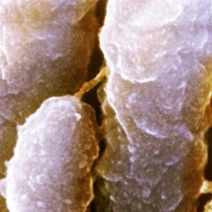
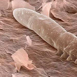
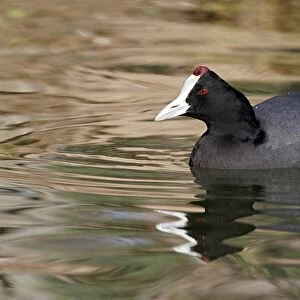
Eyelash hairs and skin. Coloured scanning electron micrograph (SEM) of eyelash hairs growing from the surface of human skin. The shafts of hair (red) are anchored in their individual hair follicles in the surface of the skin (pink). Hair is made up of a fibrous protein called keratin. The outermost skin layer, the stratum corneum, also consists of keratinized dead cells that detach from the body. The squamous (flattened) cells that make up the stratum corneum arise from the lower, living layers of skin. The tails of eyelash mites (Demodex folliculorum) are seen protruding from the base of several eyelashes. Magnification: x50 at 6x7cm size
Science Photo Library features Science and Medical images including photos and illustrations
Media ID 6455917
© STEVE GSCHMEISSNER/SCIENCE PHOTO LIBRARY
Dead Demodex Folliculorum Epidermal Epidermis Eye Lash Eye Lashes Facial Follicle Follicles Hair Hairs Histology Infection Keratin Keratinized Lash Lashes Magnified Image Microscopic Photos Mite Mites Parasite Parasites Parasitic Shaft Shafts Skin Squamous Stratum Corneum Subjects Surface Tissue Cells
FEATURES IN THESE COLLECTIONS
> Animals
> Insects
> Mites
> Follicle Mite
> Animals
> Insects
> Mites
> Related Images
EDITORS COMMENTS
This print from Science Photo Library showcases the intricate details of eyelash hairs and skin. In this magnified image, we can observe the red shafts of hair firmly anchored in their individual hair follicles on the pink surface of human skin. These hairs are composed of keratin, a fibrous protein that forms the building blocks of our hair. The outermost layer of skin, known as the stratum corneum, is also visible in this image. It consists of keratinized dead cells that naturally detach from our bodies over time. The squamous cells that make up this layer originate from the lower living layers of our skin. Interestingly, protruding from several eyelashes are tiny tails belonging to eyelash mites called Demodex folliculorum. These microscopic parasites coexist with us on our lashes without causing harm under normal circumstances. This photograph not only provides a glimpse into the anatomy and histology of healthy eyelashes but also highlights some fascinating subjects like parasitic organisms and epidermal structures. Its vibrant colors and high-resolution capture make it an excellent addition for scientific research or educational purposes.
MADE IN THE USA
Safe Shipping with 30 Day Money Back Guarantee
FREE PERSONALISATION*
We are proud to offer a range of customisation features including Personalised Captions, Color Filters and Picture Zoom Tools
SECURE PAYMENTS
We happily accept a wide range of payment options so you can pay for the things you need in the way that is most convenient for you
* Options may vary by product and licensing agreement. Zoomed Pictures can be adjusted in the Cart.