Inner ear hair cells, SEM C014 / 4844

![]()
Wall Art and Photo Gifts from Science Photo Library
Inner ear hair cells, SEM C014 / 4844
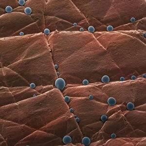
Inner ear hair cells. Coloured scanning electron micrograph (SEM) showing the sensory hair cells (blue) found in the Organ of Corti in the cochlea of the inner ear. These hairs are surrounded by a fluid called the endolymph. As sound enters the ear it causes waves to form in the endolymph, which in turn cause the hairs to move. The movement is converted into an electrical signal, which is passed to the brain. A single row of stereocilia can be seen at top, with three rows of outer hair cells below
Science Photo Library features Science and Medical images including photos and illustrations
Media ID 9224135
© CLOUDS HILL IMAGING LTD/SCIENCE PHOTO LIBRARY
Auditory Cilia Cilium Cochlea Colored Hairs Hearing Inner Ear Organ Of Corti Rows Sense Sensitive Sensory Hair Sound Stereocilia Stereocilium Surface Cells
EDITORS COMMENTS
This print showcases the intricate inner workings of our auditory system. In this coloured scanning electron micrograph (SEM), we are granted a close-up view of the inner ear hair cells, specifically those found in the Organ of Corti within the cochlea. The sensory hair cells, depicted in a striking blue hue, play a vital role in our ability to hear. Surrounded by a fluid known as endolymph, these delicate hairs respond to sound waves that enter the ear. As sound vibrations travel through the endolymph, they create ripples that cause the hairs to sway and move. This movement is then translated into electrical signals, which are transmitted to our brain for processing. The image reveals a single row of stereocilia at the top, with three rows of outer hair cells below them. Each tiny stereocilium acts as an antenna-like structure responsible for detecting different frequencies of sound. Through this mesmerizing SEM capture by CLOUDS HILL IMAGING LTD/SCIENCE PHOTO LIBRARY, we gain insight into how these remarkable biological structures function harmoniously to enable us to perceive and interpret sounds from our environment. It serves as a reminder of both the complexity and beauty inherent in human anatomy while highlighting just how sensitive and finely tuned our sense of hearing truly is.
MADE IN THE USA
Safe Shipping with 30 Day Money Back Guarantee
FREE PERSONALISATION*
We are proud to offer a range of customisation features including Personalised Captions, Color Filters and Picture Zoom Tools
SECURE PAYMENTS
We happily accept a wide range of payment options so you can pay for the things you need in the way that is most convenient for you
* Options may vary by product and licensing agreement. Zoomed Pictures can be adjusted in the Cart.