Dividing liver cancer cell, SEM

![]()
Wall Art and Photo Gifts from Science Photo Library
Dividing liver cancer cell, SEM
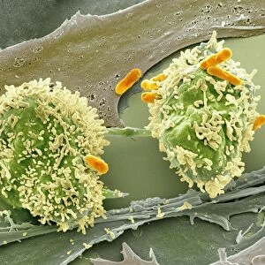
Dividing liver cancer cell. Coloured scanning electron micrograph (SEM) of a hepatocellular carcinoma (HCC) cell undergoing mitosis (nuclear division) and splitting into two daughter cells (left and right). Hepatocellular carcinoma is the most common type of liver cancer. It tends to occur in livers damaged by genetic defects, alcohol abuse, or chronic infection with diseases such as hepatitis B and C. Primary liver cancer, which starts in the liver, is relatively rare in the UK, with about 3, 600 people diagnosed each year. However, because of the prevalence of hepatitis caused by contagious viruses, it accounts for up to half of all cancers in some undeveloped countries. Magnification: x3800 when printed at 10 centimetres wide
Science Photo Library features Science and Medical images including photos and illustrations
Media ID 9222709
© STEVE GSCHMEISSNER/SCIENCE PHOTO LIBRARY
Cancer Cancerous Cell Biology Cell Cycle Colored Cytoplasmic Bridge Daughter Cells Dividing Division Filopodia Filopodium Hepatic Hepatocellular Carcinoma Hepatological Hepatology Hepatoma Liver Cancer Malignancy Malignant Mitosis Mitotic Oncological Oncology Primary Projection Projections Replicating Replication Abnormal Cells Condition Disorder Unhealthy
EDITORS COMMENTS
This print captures the intricate process of cell division within a liver cancer cell. In this coloured scanning electron micrograph (SEM), we witness a hepatocellular carcinoma (HCC) cell undergoing mitosis, where it splits into two daughter cells on the left and right sides. Hepatocellular carcinoma is the most prevalent form of liver cancer, often occurring in livers damaged by genetic defects, alcohol abuse, or chronic infections like hepatitis B and C. While primary liver cancer is relatively rare in the UK with around 3,600 diagnoses annually, it accounts for up to half of all cancers in some underdeveloped countries due to contagious viruses like hepatitis. The magnification level of x3800 allows us to appreciate the minute details present during this crucial moment in cellular replication. The image showcases various features such as cytoplasmic bridges connecting the dividing cells and filopodia projections extending from their surfaces. This visually striking representation not only serves as an educational tool for medical professionals but also highlights the complexity and abnormality associated with malignant cells. It underscores the importance of ongoing research and advancements in oncology to combat diseases like liver cancer effectively. Steve Gschmeissner's skillful capture provides valuable insight into hepatocellular carcinoma at a microscopic level while reminding us of both the challenges posed by this disease and our continuous efforts towards understanding its mechanisms for improved healthcare outcomes.
MADE IN THE USA
Safe Shipping with 30 Day Money Back Guarantee
FREE PERSONALISATION*
We are proud to offer a range of customisation features including Personalised Captions, Color Filters and Picture Zoom Tools
SECURE PAYMENTS
We happily accept a wide range of payment options so you can pay for the things you need in the way that is most convenient for you
* Options may vary by product and licensing agreement. Zoomed Pictures can be adjusted in the Cart.